BRAIN CANCER DIAGNOSIS
The first step in diagnosing brain cancer involves evaluating symptoms and taking a medical history. If there is any indication that there may be a brain tumor, various tests are done to confirm the diagnosis, including a complete neurological examination, imaging tests, and biopsy.
Imaging Tests
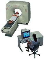
Magnetic resonance imaging (MRI scan) is the diagnostic test of choice for brain cancer. ELectromagnetic energy produces detailed computer images of the brain from different angles. It can detect edema (swelling of brain tissue) and hemorrhage (bleeding). In some cases, a dye is injected intravenously to improve the contrast between an abnormal mass and normal tissue.
Computed axial tomography (CAT or CT scan) involves the use of x-rays and a computer to obtain images of the brain. A dye is often injected intravenously to improve the contrast between an abnormal mass and normal tissue. Not only can the tumor be seen, but the type of tumor sometimes can be identified with a CT scan.
Positron emission tomography (PET scan) helps the physician evaluate brain function and cell growth by producing images of physical and chemical changes in the brain. An injected radiopharmaceutical substance is absorbed by tumor cells in the brain. Measurements of brain activity are determined by concentrations of the substance and then fed into a computer, which produces an images of the brain.
PET can precisely locate a tumor and detect metastatic and recurrent brain cancer at earlier stages than MRI or CT scan. This technique also can be used to evaluate the tumor's response to chemotherapy and radiation treatment.


